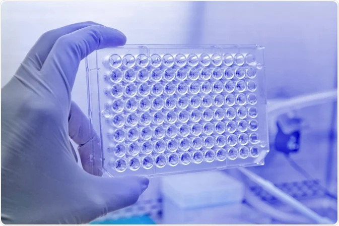
Cytotoxicity refers to the extent to which a molecule or compound can induce damage to a cell. A process or compound causing cell damage or death is called cytotoxic, where cyto means cell and toxic means poison. Cells affected with cytotoxic substances may undergo apoptosis, autophagy, necrosis, or stop actively dividing or growing to reduce cell proliferation.
Cell Cytotoxicity Assays measure the ability and extent of cytotoxic substances in damaging cells or causing cell death. Cytotoxicity screening is widely employed in drug discovery and fundamental research to screen vast libraries for toxic compounds. During drug discovery, cell toxicity is a vital endpoint for assessing the effects and fate of a candidate compound. A compound with cytotoxic effects can be appropriately eliminated from subsequent testing when evaluating the potency of pharmaceutical compounds. This article discusses cell toxicity assays, their role in drug discovery and development, and the different types of cellular and Biochemical Assays used in cytotoxicity screening.
The role of cytotoxicity screening in drug discovery and development
Drug safety is crucial in drug discovery and development. The early stages of drug formulation must incorporate wide toxicity screening of individual components, including the excipients. When a new drug candidate with promising properties enters the development process, it undergoes extensive testing to solve several complex issues.
Drug developers often employ simple yet reliable approaches to optimize the cost and efficiency of the preclinical phase of drug development. Traditional drug toxicity assessments are crucial for drug developers as safety decreases the harm to human life and financial investments. Early incorporation of cell-based assays offers a significant solution to challenges faced during human toxicity assessments. Hence, in vitro cell line systems are used to evaluate toxicity and predict human-specific harmful effects. Most importantly, the success of these Cell Toxicity Assays is due to the efforts and success of researchers and drug developers for in vitro and in vivo testing for specific absorption, distribution, metabolism, and elimination characteristics over the past decade.
In vitro systems developed to assess interaction with membrane transporters, permeability, and metabolic stability in cell models with human data have successfully reduced the failure rate to less than 10%. However, to ensure success in methods for toxicological evaluations, drug developers should employ a comprehensive and tiered screening system. Additionally, they should investigate potential limitations and liabilities of the experiments to avoid adverse data or conclusions. It is unlikely that a single in vivo or in vitro test or cell line system will suffice as a final endpoint for toxicity evaluations. Hence, researchers must conduct a well-developed toxicity screening protocol consisting of tests on multiple cell cultures.
Different types of cell toxicity assays
Enzymatic cytotoxicity assays
Glucose 6-phosphate dehydrogenase (G6PD) and lactate dehydrogenase (LDH) are enzymes present in multiple cell types. Once a cellular plasma membrane is affected or damaged, G6PD and LDH enzymes are released into the cell culture. These enzymes act as biomarkers. Researchers employ fluorescence or colorimetric methods to quantify the release of G6PD or LDH and assess cytotoxicity.
Viability assays
Researchers can detect cell viability through mechanisms such as metabolic activity, enzyme activity, and membrane integrity. Viability reagents form the foundations for an assay where cell health is evaluated through a single parameter reading or using multiple detection measures. Besides, researchers today have the option of ready-to-use kits for everyday experiments. Viability assays can assess response to external or internal stimuli, such as cell toxicity effects during drug screenings.
Apoptosis assays
Apoptosis is critical for the proper development and growth of an organism. This process eliminates excess tissues and cellular waste from infected or damaged cells. Biochemically, apoptosis causes morphological modifications such as degradation or cleavage of cellular proteins or genome fragmentation.
Autophagy assays
Autophagy is a lysosome-dependent system that recycles and degrades cellular components. Today, multiple assays are present to determine inhibition of autophagy, clearance of protein aggregates, or induction of autophagy. Lysosome markers are available to track lysosomal behaviour or fusion with autophagosomes before degradation.
Must Read: MSD Assay in Drug Development: Advantages and Use Cases
Cell proliferation assays
Cell proliferation assessments are crucial for cell differentiation, cell growth studies, and cancer research. They are widely employed to assess compound inhibition and toxicity of tumor cell growth. These evaluation markers for cell proliferation include cellular metabolism and average DNA content in a population. Cell proliferation assays can report total live cells, total cell number, or indicate DNA synthesis.
Lipotoxicity assays
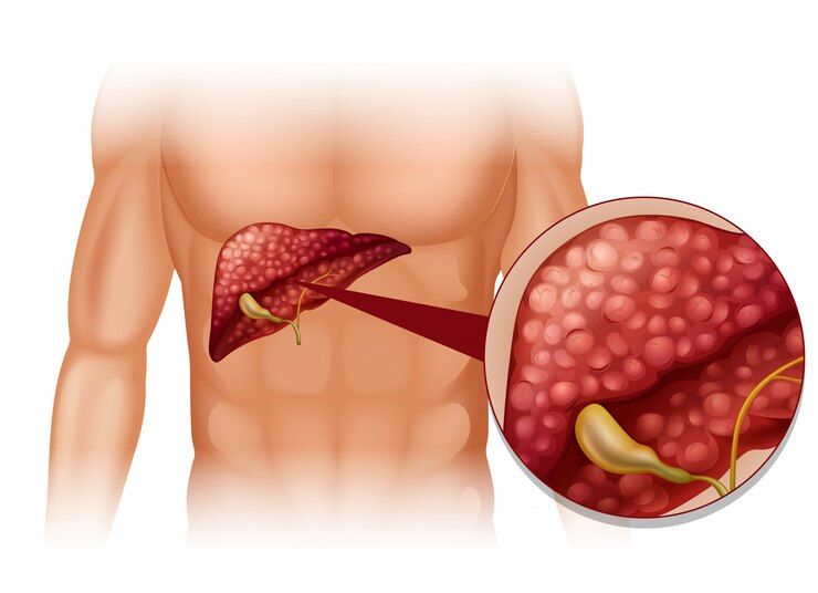
Steatosis and phospholipidosis are toxic effects of lipid metabolism. Drugs or other harmful compounds may trigger these side effects. Phospholipidosis is the accumulation of additional phospholipid complexes in the internal lysosomal membranes. On the other hand, steatosis is characterized by the retention of lipids due to abnormal elimination and synthesis of triglyceride fats. Lipotoxicity assays can measure these abnormalities.
Mitotoxicity assays
Mitochondrial functions are crucial aspects in cytotoxicity screening. These parameters are measured through calcium flux, reactive oxygen species, or mitochondrial membrane potential. Probes for mitochondrial function and structure are multiplexed to assess cellular health and address complex biological concerns on drug efficacy and cytotoxicity using microplate, imaging, or flow cytometry systems.


